
Functional Near-Infrared Spectroscopy for Assessing Executive Function in Snoring Children
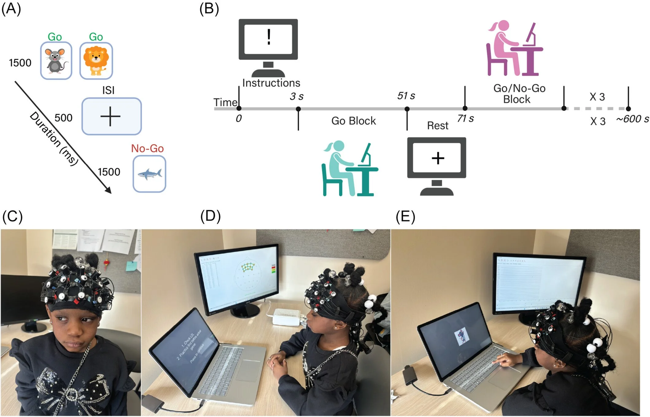
fNIRS setup for interrogating brain function in elementary school-aged children: Panel A shows the task children completed during the study. They were shown images of animals on a screen, one at a time, and asked to press a button as quickly as possible when they saw a “Go” animal, but to hold back and not press when they saw a “No-Go” animal. Each image appeared for 1,500 milliseconds, followed by a brief 500-millisecond pause before the next one. This type of task is widely used in brain research because it requires children to pay attention, stay in control of their impulses, and suppress an automatic response — exactly the cognitive skills most commonly affected in children with sleep-disordered breathing. Panel B shows how the task was structured over time. Rather than running continuously, the task alternated between blocks where children responded to every animal (Go-only blocks, used as a resting baseline) and blocks where they also had to inhibit responses to certain animals (Go/No-Go blocks, the active cognitive challenge). This alternating design allows researchers to isolate the brain activity specifically associated with the demands of attention and impulse control. Panel C shows how brain activity was recorded. Children wore a lightweight, adjustable headband fitted with sensors that used functional near-infrared spectroscopy (fNIRS) — a safe, non-invasive technique that measures changes in blood flow in the outer layer of the brain by shining harmless near-infrared light through the scalp. The headband was positioned to capture activity over the prefrontal cortex, the region at the front of the brain responsible for decision-making, attention, and behavioral control. Panels D and E show the study setup in practice — a child seated comfortably at the task display with the fNIRS headband in place, illustrating how the protocol was designed to be child-friendly and feasible even for young participants.
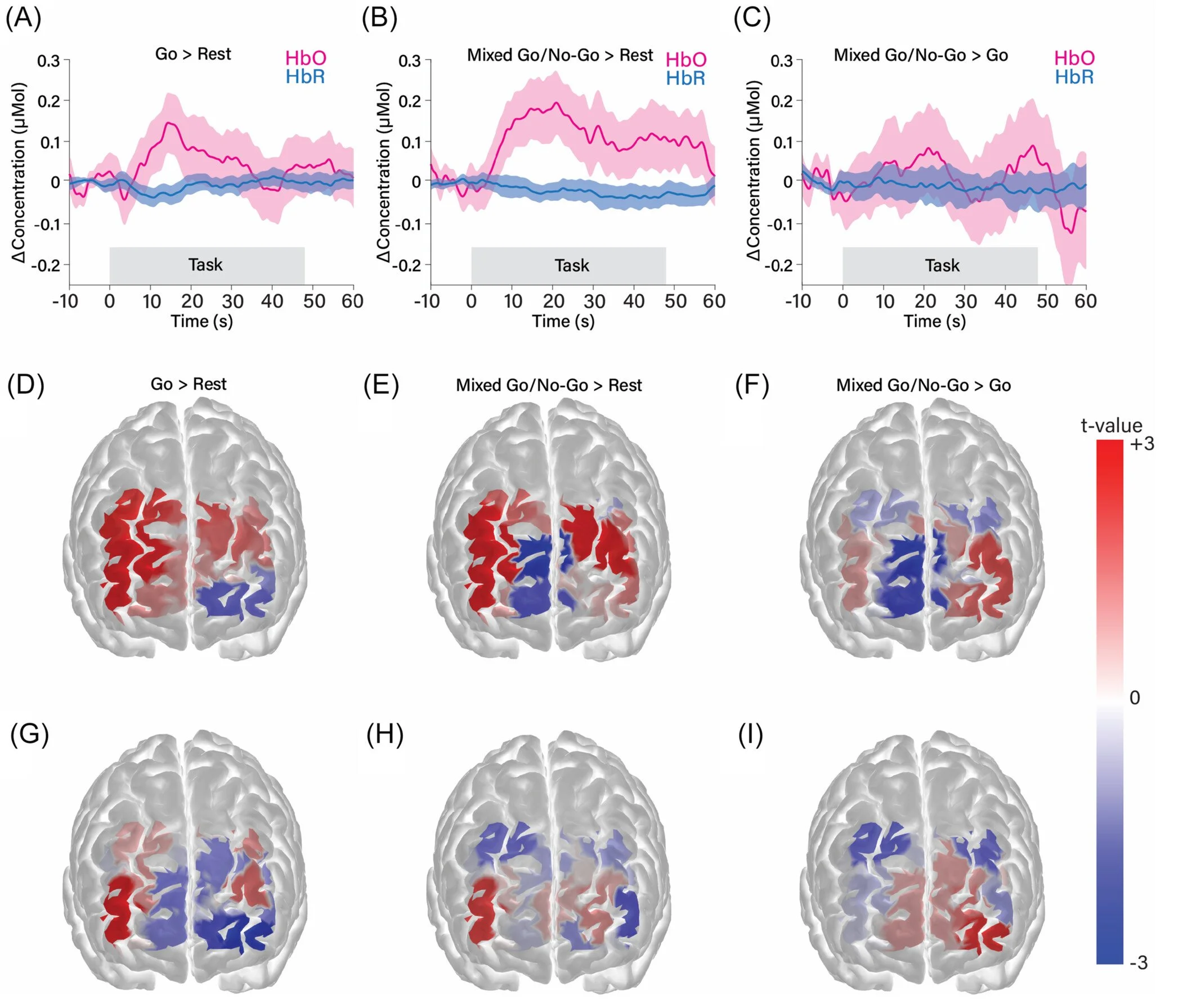
What happened in the brain during the Go/No-Go task? Panels A–C show how blood flow in the prefrontal cortex changed during different parts of the task. When the brain works harder, it draws more oxygenated blood to the active region — a signal captured by fNIRS as a rise in oxyhemoglobin (HbO, the oxygen-carrying form of hemoglobin) and a corresponding dip in deoxyhemoglobin (HbR, the oxygen-depleted form). These curves, plotted over time, are called hemodynamic responses and serve as a proxy for neural activity. Panel A shows the brain's response during the simple Go blocks compared to rest — reflecting the baseline demands of paying attention and pressing a button. Panel B shows the response during the mixed Go/No-Go blocks compared to rest, which captures the combined demands of attention and impulse control. Panel C is the most informative comparison: the Go/No-Go blocks directly contrasted against the Go-only blocks. By subtracting out the shared demands of basic attention, this comparison isolates the brain activity specifically required to suppress a response — the cognitive act of holding back — which is the core executive function tested by this task. Panels D–I translate these signals into maps of the prefrontal cortex, showing which specific regions were most active during each comparison. The maps are shown separately for HbO (panels D–F, where warmer colors indicate greater oxygenation) and HbR (panels G–I, where the expected pattern is a mirror image — a decrease in the same regions). Together, these two signals provide converging evidence for where in the prefrontal cortex the brain is doing the extra work of inhibitory control — and lay the foundation for comparing that activation between children with and without sleep-disordered breathing.